Brain Aneurysm / Cerebral Aneurysm

Dr Newell has devoted his career to the management and treatment of patients with cerebral aneurysms. He is also a recognized national and international expert on the treatment of cerebral aneurysms and has authored and co-authored multiple articles and several textbooks on the care and treatment of patients with brain aneurysms.  Having operated on over 1,500 brain aneurysms, Dr Newell has vast surgical experience in surgical treatment and endovascular and critical care management of patients with cerebral aneurysms. He has also trained a large number of neurosurgeons seeking advanced surgical skills on surgical techniques to treat complicated cerebral aneruysms. 

Although many people think a brain aneurysm or cerebral aneurysm means sudden, unexpected death, this condition can be both survivable and treatable. Neurosurgeons who are specially trained in cerebrovascular diseases, evaluate most patients with aneurysms. Some patients with cerebral aneurysms, have them discovered on imaging studies such as MRI or CT done for headache pain, dizzyness, or other reasons. More definitive studies to characterize aneurysms include computerized tomography angiography (CTA), magnetic resonance angiography (MRA), and cerebral angiography. The risk of an aneurysm rupturing includes many factors related to the aneurysm itself including size, location, anatomic features, as well as patient factors including genetics. Many treatment options exist now to safely eliminate aneurysms from the circulation and prevent rupture and pressure symptoms from the aneurysms.

PDF: Cerebral Aneurysms

If you need help with any of these conditions, please contact us to get help!

Or call 206.661.6100 for immediate assistance

What is a brain aneurysm?

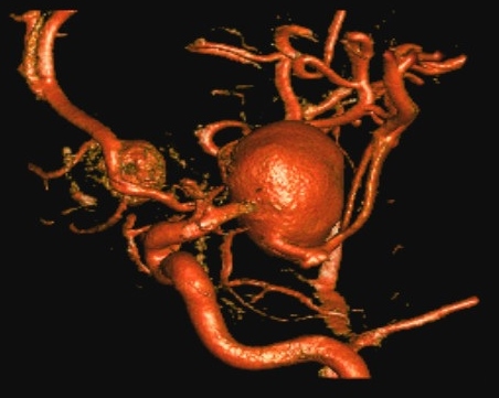
A brain aneurysm (also called a cerebral aneurysm) is a blister-like bulge in a weak part of a blood vessel in your brain. It can go unnoticed for a long time. In fact, 1-6 percent of Americans has an aneurysm they don’t know about. If left untreated, however, the pressure of the blood causes that area of wall of the blood vessel to become even weaker, which allows the aneurysm to grow. Eventually the aneurysm may burst, which causes a stroke.

What do I need to know?

A few simple rules will help you avoid a brain aneurysm and/or help you receive essential and timely treatment.

  • Know the risk factors and create a lifestyle that helps you avoid an aneurysm in the first place
  • If you are at risk for brain aneurysm, talk with your doctor about getting screened for early detection
  • If you experience any of the symptoms,call 911 so you can receive treatment right away

Where in the brain do aneurysms develop?

The internal carotid arteries are the two large arteries – located on either side of the head and neck – that supply blood to the brain.

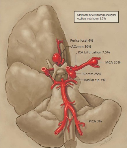
The most common types of aneurysms are found in the arteries that connect the two internal carotid arteries:

  • 30% of aneurysms form in the anterior communicating artery (AComm)
  • 25% form in the posterior communicating artery (PComm)

The frequency of aneurysms in other locations:

  • 20% in the middle cerebral artery (MCA), which supply blood to the front of the brain
  • 7.5% in the internal carotid arteries themselves, called ICA bifurcation
  • 7% in the tip of the basilar artery, which runs along the middle of the brain, where it divides into two smaller arteries
  • 4% in the pericallosal artery, which is a segment of another important pair of arteries called the anterior carotid arteries
  • 3% in the posterior inferior cerebellar artery (PICA), which supplies blood to the bottom portion of the brain

A small percentage of aneurysms form in other areas of the brain as well.

Symptoms

Whether or not you are at risk for brain aneurysm, it is wise to know the signs and symptoms.

At first, you may not know you have a brain aneurysm because it may be present for a long time without causing any symptoms. It may be discovered only when you have testing or imaging for some other condition. That is why it is important for you to talk with your doctor about early detection if you are at risk for aneurysm.


Signs and symptoms that an aneurysm is growing

An aneurysm that is growing, but hasn’t burst, may cause pressure on nerves or tissue in the brain. This pressure may cause:

  • Headache or neck pain
  • Fuzzy or double vision, or loss of vision
  • Numbness in the face
  • Enlarged pupil size
  • Drooping eye lid
  • Severe drowsiness


Signs and symptoms that an aneurysm has burst

An aneurysm that has burst or ruptured allows blood to flow into the space surrounding the brain (subarachnoid space) or directly into the brain. The symptoms of a ruptured aneurysm come on suddenly. You may experience:

  • A sudden, severe headache (many people describe this type of headache as “the worst headache of my life”)
  • Severe stiff neck
  • Nausea and vomiting
  • Double vision
  • Mental confusion
  • Dizziness and fainting
  • Losing consciousness

If you – or a friend or relative – experience any of these symptoms, call 911 immediately.

Risk Factors

Anyone can develop a brain aneurysm at any time. About one in 40 people will develop a brain aneurysm, usually between the ages of 40 and 60.


Aneurysm risk factors you cannot control:

Sometimes a brain aneurysm is caused by conditions that you cannot control, such as:

  • Family history: If two or more close relatives (parents, brothers or sisters, or children) have aneurysms, you may be at risk for a “familial aneurysm.” This type of aneurysm tends to rupture while it is still small
  • Birth defect: You may have been born with a defect in one of your blood vessels that allows an aneurysm to develop
  • Gender: Women are more likely than men to develop a brain aneurysm
  • Race: African Americans are more susceptible to brain aneurysm
  • Disease, infection, circulatory disorder: Some genetic diseases, and some types of infections and circulatory disorders may cause a brain aneurysm. Aneurysms that occur after an infection in the heart or blood vessels are called mycotic aneurysms
  • Trauma: An accident with trauma to the head may cause a traumatic aneurysm, a rare type of aneurysm in an artery in the brain

If you have any of these risk factors, you should talk with your doctor about your family and personal medical history, and ask about early detection screening.


Aneurysm risk factors you can control:

There also are many risk factors you can control or manage with lifestyle changes. These include:

  • Smoking: If you smoke, STOP. Smoking has been associated with the formation, growth and increased rate of rupture of aneurysms
  • Alcohol abuse: If you drink, only drink in moderation. If you have any genetic risk factors for aneurysm, consider avoiding alcohol completely
  • High blood pressure (hypertension): Help control your blood pressure through diet and exercise, and/or medication if your doctor recommends it
  • Drug abuse: Avoid recreational drug use – especially cocaine


If you are at risk for a brain aneurysm, make the necessary lifestyle changes and talk with your doctor about early screening.

Diagnosis

Neurosurgeons use a variety of imaging tests to diagnose a brain aneurysm. You may have one or more of the following imaging exams:

CT Scan and CT Angiography (CTA): A CT scan of the brain is often the first test your neurosurgeon will request. A CT scanner uses X-rays to create images of very thin sections of your brain that help identify the location and size of an aneurysm. It also helps determine if there is blood leaking from the aneurysm. A CTA is a CT scan taken after a small amount of dye is injected. The CTA produces images of the blood vessels and can show aneurysms.

MRI or MRI Angiography (MRA): An MRI or MRA are additional methods of determining the location, size and condition of an aneurysm, and whether the aneurysm is bleeding into the skull. MRI and MRA use magnets, radio-wave energy and computers to create images. The images can be either 3-D or 2-D. MRI and MRA are very similar, except an MRA uses a small amount of dye to help highlight the aneurysm.

Cerebral Angiogram: The neurosurgeon will use a cerebral (brain) angiogram to see how the blood is flowing through the arteries and veins in your brain. During this X-ray test, a small amount of dye is injected through a tiny tube (catheter) into the blood vessel in the brain to make it easier to see the blood flow. The angiogram can identify defects in a blood vessel, such as a blockage or a weak area that may develop into an aneurysm. It is often used to detect very small aneurysms.

Lumbar Puncture: Your doctor may request a lumbar puncture if he or she suspects your aneurysm is bleeding into your brain. A needle is inserted into the lower-back portion of the spine to withdraw a small amount of spinal fluid. The fluid is examined to determine if there is blood present.

For Medical Professionals

Brain aneurysms are abnormal dilatations or outpunching of cerebral vessels which can cause a number of different symptoms in patients resulting from pressure effects on nerves or surrounding tissue and can also place patients at risk for hemorrhage in the brain with serious consequences.

Types of Aneurysms

There are a number of types of aneurysms which affect the brain and may behave quite differently depending on the type, size, location, shape of each aneurysm and whether they have caused symptoms or not. Most aneurysms result from the disruption of the normal structure of the cerebral blood vessels which can occur from various inherited factors, or from factors which effect patients during their life and cause damage to the vessels resulting in aneurysm formation.

The major types of brain aneurysms include:

  • Saccular or Berry aneurysms
  • Fusiform aneurysms
  • Traumatic aneurysms
  • Infectious or Mycotic aneurysms

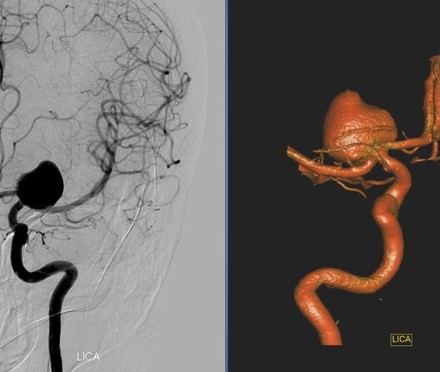
Saccular, or berry aneurysms are the most common types of aneurysms, and usually occur at predictable locations along the cerebral vasculature. They resemble a small berry or sac, and project off of the side of cerebral vessels, usually at branch points.

These aneurysms may also be referred to in the medical or healthcare literature as “congenital aneurysms” or “congenital berry aneurysms”, however these terms are based on information that is now known not to be accurate when it was believed that patients were born with aneurysms and they only ruptured later in life. In the past, before modern imaging techniques including magnetic resonance angiography (MRA) or computerized tomography angiography (CTA), aneurysms were only diagnosed with cerebral angiography, generally after they ruptured or caused symptoms and therefore were often not detected before rupture.

It is rare to find aneurysms in infants, children or adolescents, however they do occur in these age groups on occasion on a very infrequent basis. Saccular aneurysms are thought to arise from focal defects in the blood vessel wall combined with sheer stresses caused by the pulsation jets and turbulence which results from the blood flow within the vessels as it reaches branch points and significant angles and direction shifts.

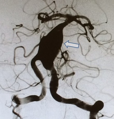
Fusiform brain aneurysms are dilatations of the cerebral vessels that result in the loss of the normal tubular structure of the vessel and the appearance of significant irregular sac-like widening and sometimes elongation of the vessels. Fusiform aneurysm also are believed to be caused by structural defects in the blood vessel wall which can be caused by dissection, or tearing of the internal lining of the vessel wall resulting in the blood flow entering the wall of the blood vessel resulting in and enlargement of the space where the blood flow is contained.

Fusiform aneurysms can arise in patients with a normal vasculature, who may have environmentally induced stressful insults to the vessels, and also can arise with patients with collagen (fibrous material that holds body tissue together) vascular disease, who have genetic abnormalities which prevents them from forming and maintaining the normal structural elements of the blood vessel. Weakening of the structure of the blood vessels from a variety of causes can result in the blood pressure inside the vessel forcing the vessel wall apart to form fusiform aneurysms which resemble sac or sausage like dilatations of the vessels.

Traumatic aneurysms are rare and usually occur only after a vessel is directly injured by trauma. Head trauma is often classified as either closed head injury, where the covering of the brain is not violated, or as penetrating injury, where an object has penetrated through the covering of the brain and can directly injure the brain tissues and also the brain blood vessels.

Examples of penetrating injury include gunshot wounds to the head, stab wounds to the head, or compound depressed skull fractures where bone fragments can be driven into the brain and cause direct injury. Traumatic aneurysms are almost exclusively seen after penetrating injury and rarely occur following closed head injury. It is believed that direct injury to the vessel wall results in out pouching of the inner lumen or inside tubular structure of the vessel to cause the formation of traumatic aneurysms.

Infectious or mycotic aneurysms are most commonly seen after a blood stream infection where microorganisms are growing inside the vessel walls. This situation can occur with intravascular infections from a variety of causes, however the most common setting in which mycotic brain aneurysms are seen is in subacute bacterial endocarditis.

In this condition bacteria begin to grow and form vegetations, or small outgrowths of tissue on the leaflets of the heart valves. These vegetations or outgrowths can break off into the bloodstream and travel up into the cerebral circulation where they can lodge in the more peripherally located brain vessels. The bacteria can weaken the vessel wall and the vessel dilates and becomes a mycotic aneurysm.


Signs and Symptoms & Discovery on Imaging Studies

Signs and symptoms of brain aneurysms fall into several categories. The most common signs and symptoms are either from direct pressure effects from enlarging aneurysms on either the cranial nerves or other brain structures, or preparing to rupture resulting in a so called “warning leak”. Occasionally larger aneurysms can undergo blood clot formation within them and cause transient ischemic attacks (warning signs of stroke), or actual strokes. In rare circumstances larger aneurysms can cause seizures by direct pressure effect on the brain.

In many cases, patients may present to medical attention with neurologic symptoms which are of uncertain significance, or symptoms such as light headedness or dizziness which may have nothing to do with an aneurysm, and an aneurysm may be discovered on either an MRI or CT which is ordered to evaluate the symptoms. If an aneurysm is found on an MRI or CT then more sensitive test called MRA, or magnetic resonance angiography, or CTA, computerized tomography angiography can be performed.

The gold standard test for detecting and defining the critical features of cerebral aneurysms is cerebral angiography where contrast material is directly injected in the proximal cerebral arteries through a transfemoral approach through the leg. X-rays are taken of the head with contrast material in the brain blood vessels, and 3D spin angiography is used to complement the x-ray images by performing computerized reconstructions of images obtained by filming the vascular structures as the x-ray tube is spun around in an arc around the patient.


Subarachnoid Hemorrhage

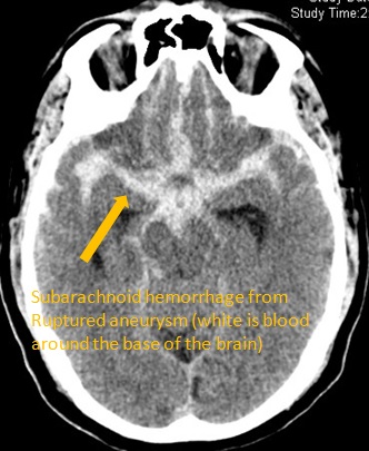
Subarachnoid hemorrhage (SAH) refers to bleeding within the brain in the subarachnoid space, which is the space where the cerebrospinal fluid (CSF) circulates, over the surface of the brain but under the arachnoid membrane which is a cellophane like semi clear membrane that covers the brain and cerebral blood vessels. Cerebral aneurysms are the most frequent cause of spontaneous SAH, with arteriovenous malformations accounting for approximately 6%, Other causes of SAH include trauma, tumors, and vasculitis (inflammation of the blood vessels).

Many retrospective studies, which record symptoms in groups of patients that have aneurysms rupture, suggest that up to 40% of patients have “warning leaks” though some investigators would argue the true incidence is lower. Most occur 1–8 weeks before a major SAH and may not necessarily be seen on CT or lumbar puncture. Headache is usually milder but of similar nature and sudden-onset, lasting several days, and can be associated with nausea and vomiting, although stiff neck and light sensitivity are rare.

Patients who have experienced an SAH usually complain of an acute onset terrible headache described as a thunderclap headache, or the “worst headache of my life.” The headache is usually persistent, and is often accompanied by stiff neck and light sensitivity. SAH is often preceded in time (usually days or weeks) by a less severe headache, also of sudden onset, that clears within a 24 hour period and is referred to as a “warning headache” or “sentinel hemorrhage.” This event is thought to represent a small microscopic leak, or leak within the vessel wall from the aneurysm and a precursor to the full blown SAH.

A non-contrast head CT scan is the diagnostic test of choice to confirm the diagnosis of SAH. If performed within 24 hours of the SAH, the CT should be able to detect 90-95% of all SAH. The blood from SAH is usually located in the basilar cisterns, extending into the sylvian and/or interhemispheric fissure of the brain and can be accompanied by with variable amounts of intraventricular or intracerebral blood.

Lumbar puncture is reserved for patients who have a normal head CT despite a story suggestive of an SAH. Common reasons for a CT scan to be negative following SAH are, very minor hemorrhage, prolonged interval of over several days between the headache and the CT scan which may allow blood to diffuse away from the bleeding site and not be readily detectable on CT. Appropriate interpretation of the results of the cerebrospinal fluid obtained from lumbar puncture can make the diagnosis of SAH.


Treatment of Brain Aneurysms

There are four main options for treating patients with brain aneurysms: observation, craniotomy with clip ligation (“clipping”) and endovascular occlusion using detachable coils (“coiling”), or parent vessel ligation where the vessel leading up to an aneurysm is ligated or occluded. This can be done with either a clip surgically, or using an endovascular device.

All ruptured brain aneurysms are treated in patients with a reasonable chance of surviving, whereas incidentally discovered unruptured aneurysms may be either treated of observed depending on aneurysm and patient factors including aneurysm size and location and patient age and general health. Observation includes routine periodic imaging and physician visits where these studies are reviewed.

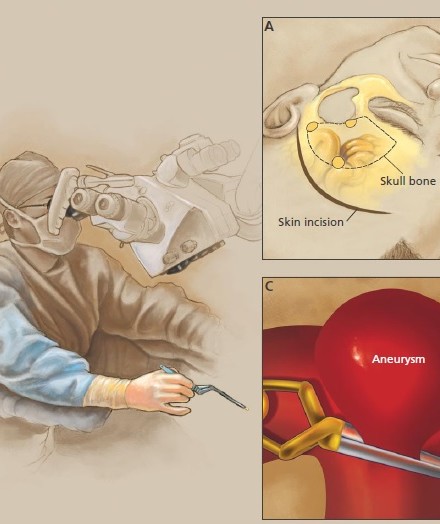
Clipping of aneurysms requires open surgery using craniotomies performed by neurosurgeons under general anesthesia. The operating microscope is used to expose the aneurysm and allow safe clip placement. Permanent clips made from metals that are MRI-compatible are placed across the neck of the aneurysm, excluding it from the circulation.

Endovascular occlusion or coiling is performed by either an interventional neuroradiologist, or a neurosurgeon or neurologist with interventional training. During coiling, the patient is most
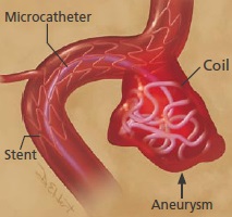
often placed under general anesthesia, though this is not always the case. Angiography is performed following which a microcatheter is navigated into the aneurysm and detachable coils of varying sizes and shapes are deployed within the aneurysm to decrease or eliminate filling of the aneurysm with blood. More recently intracranial stents have been utilized as an adjunct to coiling to prevent the coils from prolapsing into the parent vessel in aneurysms which have a wide neck.


Related medical articles and books

LeRoux PD, Newell DW, Winn HR: Management of Cerebral Aneurysms; Saunders, Philadephia, Pennsylvania. 2004

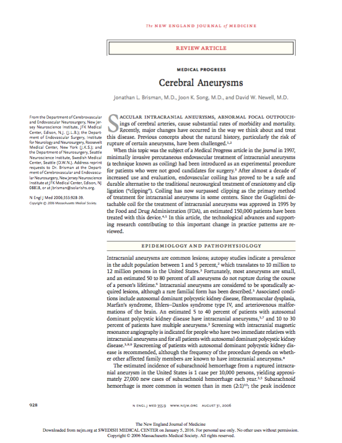
Brisman JL, Song JK, Newell DW: Cerebral aneurysms. N Engl J Med. 2006 Aug 31;355(9):928-39. Review

Britz GW, Salem L, Newell DW, Eskridge J, Flum DR: Impact of Surgical Clipping on Survival in Unruptured and Ruptured Cerebral Aneurysms –A Population-Based Study. Stroke, Vol, 35, No. 6, June 2004, pp.1399-1403

Newell DW, Avellino A, Schuster J: Intracranial to intracranial vascular anastomosis using the microanastomotic device for the treatment of distal middle cerebral artery aneurysms. J of Neurosurgery v 97: 2002; 20-25

Elliott JP, Le Roux PD, Ransom G, Newell DW, Grady MS, Winn HR: Predicting length of hospital stay and cost by aneurysm grade on admission. J Neurosurg. 1996;85:388-391

Le Roux PD, Elliott JP, Newell DW, Grady MS, Winn HR: The incidence of surgical complications is similar in good and poor grade patients undergoing repair of ruptured anterior circulation aneurysms: A retrospective review of 355 patients.Neurosurgery. 1996;38:887-895

Le Roux PD, Elliott JP, Newell DW, Grady MS, Winn HR: Predicting outcome in poor grade subarachnoid hemorrhage: A retrospective review of 159 aggressively managed patients. J Neurosurg. 1996;85:39-49

Le Roux PD, Elliott JP, Downey L, Newell DW, Grady MS, Mayberg MR, Eskridge JM, Winn HR: Improved outcome after rupture of anterior circulation aneurysms: a retrospective 10-year review of 224 good-grade patients.J Neurosurg.1995;83:394-402

Le Roux PD, Grady MS, Newell DW, Dailey D, Winn HR: Emergent aneurysm clipping without angiography in the moribund patient with intracerebral hemorrhage:The use of infusion computed tomography scans. Neurosurgery. 1993;33:189-197

Eng CC, Lam AM, Byrd S, Newell DW: The diagnosis and management of a perianesthetic cerebral aneurysmal rupture aided with transcranial Doppler ultrasonography. Anesthesiology. 1993;78:191-194

Newell DW, Eskridge J, Mayberg M, Grady MS, Lewis D, Winn HR: Endovascular treatment of intracranial aneurysms and cerebral vasospasm. In ClinicalNeurosurgery, Selman W (ed), Williams & Wilkins, Baltimore. 1992:p348-360

Newell DW, Eskridge JM, Mayberg MR, Grady MS, Winn HR: Angioplasty for the treatment of symptomatic vasospasm following subarachnoid hemorrhage. J. Neurosurg.1989;71:654-660


Treatment

Brain aneurysms are not always treated immediately. Sometimes, if the aneurysm is very small and is not leaking, your doctor may prefer to monitor it for awhile, especially if there are other medical conditions that need to be managed.

Your doctor may recommend imaging every year or on a regular basis, to observe the aneurysm and determine if it is growing.

When it is time to treat the aneurysm, there are four main options:

  • Surgical clipping
  • Parent vessel occlusion (ligation)
  • Coiling (embolization)
  • Pipeline® Device (flow diversion)

The brain aneurysm team at Swedish has many years of experience in all of these treatment options, and in protecting patients from the risk of stroke following treatment.

Clipping

This procedure requires the neurosurgeon to enter the brain to place a small clothes-pin-like clip around the base of the aneurysm where it protrudes from the blood vessel. The clip blocks the blood flow to the aneurysm so there is no more pressure applied to the weakened wall of the blood vessel. The blood that remains in the aneurysm clots, and there is no longer a threat the aneurysm will burst and bleed into the skull.

Parent Vessel Occlusion

Sometimes an aneurysm has damaged the blood vessel so much that the neurosurgeon must clamp off the entire artery. This is a more complicated surgery that may also require a bypass to re-route the blood around the clamped section of the vessel. The choice of treatment is primarily determined by the size, shape and location of the aneurysm, as well as your medical condition, overall health and family history.

Coiling

Coiling is a minimally invasive procedure used to repair the aneurysm from inside the blood vessel. An interventional neuroradiologist threads a very thin, hollow tube (micro catheter) through the blood vessel to the location of the aneurysm. This tube is used to transport a tiny, tightly coiled bundle of platinum fibers to the aneurysm.

While viewing the procedure on a monitor, the surgeon inserts the coil into the aneurysm to protect the weakened wall of the blood vessel from the pressure of the blood flow. Sometimes a tiny splint-like sheath (stent) is inserted into the blood vessel to help strengthen that section of the vessel and to help keep the coil in place. 

Flow diversion and Vascular reconstuction

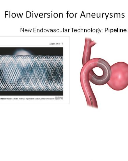
Flow diversion is an endovascular procedure that can be used to treat large or giant wide-neck brain aneurysms in adults. The devices used for flow diversion are similar to stents only the have a much tighter woven metal structure in which the metal covers a larger surface area than a standard intracranial stent. The tighter weave provides a more significant barrier to flow than a standard stent and restricts the flow going into an aneurysm and ideally results in slow flow and delayed trombosis of the aneurysm. Pipeline is one of the first brands that was approved for use.

The Pipeline® Device restores original, natural blood circulation while providing permanent long-term occlusion. During the procedure, the device (a braided cylindrical mesh) is implanted across the aneurysm neck. This slows the flow of blood into the aneurysm, which allows the diseased vessel to heal.

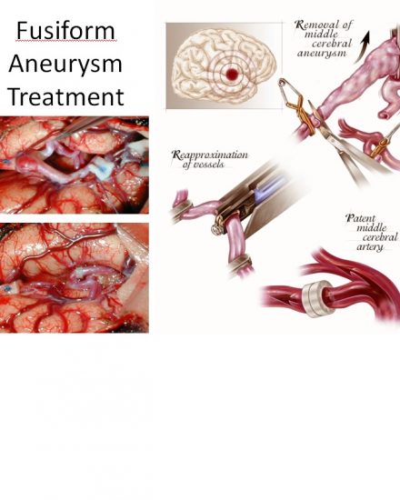
Vascular reconstruction can be used for complex aneurysms which cannot be treated with either standard clipping or using endovascular techniques. Vascular reconstruction can include extracranial to intracranial bypass or intracranial to intracranial bypass as shown below. These techniques are especially useful in treating large complex and fusiform aneurysms.

Video

In this video, Dr. David Newell defines and explains what a cerebral aneurysm is. A cerebral aneurysm is a weak spot on one of the blood vessels that goes into the brain, and one to two percent of all adults have aneurysms in the cerebral circulation. Dr. Newell shows where aneurysms can be located, and discusses the risks involved when an aneurysm ruptures.  The first video covers all the  frequently asked questions about cerebral aneurysms.  The next series of 8 videos is the first video broken down into 8 parts with easy access to each frequently asked question.

Frequently asked questions

Cerebral aneurysms: what are they?

Frequently asked questions

Cerebral aneurysms: How are they discovered?

Cerebral aneurysms

What are the signs and symptoms?

Cerebral aneurysms

Are they something you are born with?

Cerebral aneurysms

How are they treated?

Cerebral aneurysms

What are the success rates for clipping or coiling?

Cerebral aneurysms

What is the bottom line?

Cerebral aneurysms

Parting thoughts and recommendations

Brain Aneurysm | Cerebral Aneurysm. For help with symptoms, risks, and treatment options, contact our office.

Dr. David W Newell was born in Boston, MA and attended Case Western Reserve University Medical School. He completed his residency in neurosurgery at the University of Washington, including a year in London at St. George’s Medical School. Dr. Newell is the co-founder of the Swedish Neuroscience Institute and founder of the Seattle Neuroscience Institute.