For an appointment about pain, symptoms, risks, and treatment contact us or to learn more about your world renown surgeons at Seattle Neuroscience Institute visit our home page.
Seattle Neuroscience Institute
Spinal Canal and Spinal Cord Tumors
Spinal tumors are broadly classified into tumors which are located in or arise from the vertebral and bony elements of the spine, and those which occur in the spinal canal. They can also be referred to according to the regions of the spine in which they are located which are termed cervical, thoracic, lumbar, and sacral. Tumors of the vertebral column, or bony elements include both benign and malignant tumors arising from the bone elements and cartilage, or metast atic tumors which spread to the bone from other sides including blood elements such as myeloma or from a variety of other organs and tissues including prostate, breast, kidney, thyroid, lung, and other sites.
Spinal canal tumors can arise from the tissues covering the spinal cord and are called either exdradural or intradural depending on whether they are on the outside or the inside of the dural membrane which contains the spinal fluid, nerve roots and spinal cord. The intradural tumors are further broken down in to those that are inside the dura but outside the spinal cord, and are referred to as intradural extra-medullary tumors, and those which are inside the spinal cord and are referred to as intradural intra-medullary tumors. The most common types of tumors which are found in the spinal canal, outside the spinal cord are meningiomas and schwannomas and metastatic tumors which most commonly extend from the bone to enter the spinal canal.
Symptoms
- Progressive weakness of the legs
- Progressive weakness of the arms and hands
- Difficulty walking and stumbling
- Problems with bowel or bladder function
- Altered sensation on one side of the body below the neck or in the arms or legs
Causes
Tumors of the spinal canal and spinal cord are usually benign but can also be malignant and may occur as a result of over growth of a cell type that is usually found in the spinal cord tissue. Common types of spinal cord tumors include:
- Ependymoma; a tumor arising from the ependymal cells that line the central canal of the spinal cord or arising in the filum terminale which is a fibrous cord-like structure that comes off the lower end of the spinal cord
- Astrocytoma; a tumor which arises from the astrocytes or supporting cells of the spinal cord
- Hemangioblastoma; a tumor which arises from the blood supporting cells of the spinal cord and may be associated with cysts. These tumors may occur alone or may be multiple in the spinal cord and brain and can occur with Von Hippel Lindau disease, which may have a genetic origin
- Meningiomas; a tumor which arises from the covering of the spinal cord and can exert direct pressure on the spinal cord and nerve roots
- Schwannoma; schwannoma of the spinal canal usually arises from the dorsal or posterior part of one of the nerve roots and can grow large enough to deform the spinal cord and exert direct pressure on it
Risk Factors
Most spinal canal and spinal cord tumors occur sporadically and do not have particular risk factors. Hemangioblastomas of the spinal cord are a known risk factor in patients with Von Hippel Lindau disease.
Diagnosis
Your doctor will ask about your symptoms and medical history. A physical exam will be done. Tests may include:
- MRI scan of the spinal canal and spinal cord with and without contrast material
- Angiography —uses contrast material to see blood vessels
- Plain x-rays and CT scan of the spinal canal
Prevention
There are no current guidelines to prevent the formation of spinal tumors.
Treatment
Talk with your doctor about the best treatment plan for you. Options include:
The approach to spinal tumors is to obtain a working diagnosis to avoid surgery when not needed and employ safe surgical techniques with appropriate neuromonitoring and intraoperative adjuncts to achieve the best result. Patients should have a thorough investigation and work up when they are found to have a spinal abnormality to make sure a tumor diagnosis is not made of the patient has other conditions which can mimic spinal cord tumors such as multiple sclerosis, inflammation, infarction (stroke) of the spinal cord, vascular fistula, or cavernous malformation of the spinal cord.
When surgical treatment is indicated patients will obtain diagnostic studies to properly pinpoint the location of the tumor and plan the extent of the exposure to the tumor which is a laminectomy through a posterior spinal approach in most cases.
Surgical Removal
Surgical removal of a cervical spinal cord ependymoma using a CO2 Laser. Ependymomas are usually benign tumors of the spinal cord which generally have a good prognosis if they can be completely resected. Like some hemangioblastomas, epenymomas can form a cyst in the spinal cord above and or below the tumor. Figure 3 shows the spinal cord (a) before, (b) during, and (c) after the removal using a CO2 Laser under a high powered microscope.
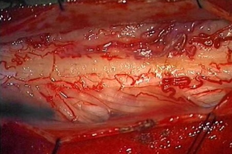 Figure 3a
 Figure 3b
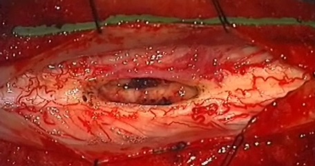 Figure 3c
Laser Removal
Laser removal of lumbar schwannoma. Tumors in the intradural space in the lumbar region are most commonly either schwannomas or ependymomas attached to the filum terminale which is a fibrous and vascular structure that comes off of the lower end of the spinal cord. Figure 4 shows a schwannoma (indicated by star) of the lumbar region (a) before, (b) during, and (c) after removal with a CO2 Laser.
 Figure 4a
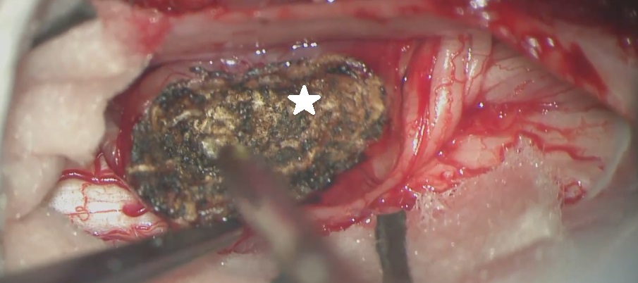 Figure 4b
 Figure 4c
Case Examples
Hemangioblastoma of the spinal cord. Hemangioblastomas of the spinal cord are benign tumors which can come to medical attention due to progressive growth and pressure on the spinal cord which can produce symptoms of weakness or numbness of the limbs. These tumors can also sometimes cause a cyst in the spinal cord which is associated with it and usually disappears when the tumor is removed. The surgical removal is performed after a laminectomy and opening of the dura mater which covers the spinal cord and contains spinal fluid. The tumor removal is performed under a high powered microscope using bipolar cautery to shrink the tumor and remove it.
 Figure 1a
 Figure 1b
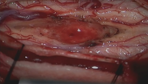 Figure 1c
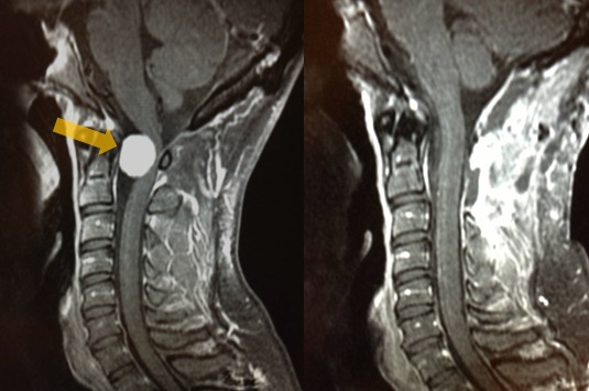
Meningioma of the upper spinal canal. Meningiomas of the spinal canal are most always benign tumors and produce pressure effects on the spinal cord usually causing numbness and weakness which is often slow in onset and progressive in nature. The preferred imaging study is an MRI with and without contrast which shows the tumor very clearly in most cases Figure 2a (left, before surgery) (right, after surgery). The tumor is removed by opening the dura, which covers the spinal cord over the extent of the tumor, to gain access to the upper and lower portion of the tumor. Figure 2b shows tumor to the left (star) and spinal cord to the right with nerve roots crossing behind the tumor before removal. Figure 2c shows the spinal cord to the right after tumor removal.
 Figure 2a
 Figure 2b
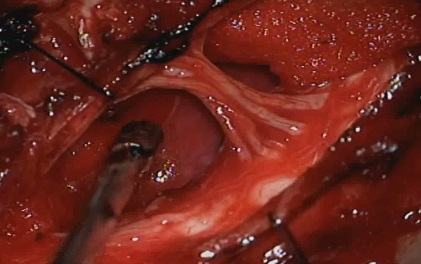 Figure 2c
Video
This video shows the microsurgical removal of a tumor inside the spinal cord called a hemangioblastoma with small associated cyst.
Warning: this video contains graphic depictions of neurosurgery.
Cervical spinal cord ependymoma removed with microsurgical techniques and CO2 Laser. Surgery performed by David W Newell MD
Warning: this video contains graphic depictions of neurosurgery.