Cavernous Malformations

Cavernous malformations are clusters of abnormal, tiny blood vessels, and larger, stretched-out, thin-walled blood vessels filled with blood in the brain. These blood vessel malformations can also occur in the spinal cord, the covering of the brain (dura), or the nerves of the skull.

Symptoms

  • Repeated severe headaches
  • Seizures or epilepsy
  • Bleeding in the brain
  • Repeated bleeding episodes in the brain
  • Progressive neurological deficits including motor weakness, sensory disturbance or
    cranial nerve problems

Causes

Cavernous malformations can arise spontaneously in the brain or spinal cord and most commonly occur in adults although approximately 25% of all diagnosed cavernous malformations are found in children
 
  • Most cavernous malformations occur randomly, so called sporadic cavernous malformations
  • Some cavernous malformations can be genetic in origin and run in families, so called familial cavernous malformations.  Familial cavernous malformations accounts for at least 20% of all cases and follows autosomal dominant inheritance. Individuals with familial CCM have a 50% chance of passing the illness to each child
  • Some cavernous malformations can be multiple and occur in many locations in the brain and spinal cord

Risk Factors

Genetic predisposition in the familial type.

Diagnosis

Your doctor will ask about your symptoms and medical history. A physical exam will be done. Tests may include:
 
  • CT scans
  • Angiography — uses contrast material to see blood vessels to rule out arteriovenous malformation and to look for venous malformations which may co-exist with cavernous malformations
  • MRI scan of the brain and or spinal cord

CT scan of cavernous malformation:

MRI scan of cavernous malformation (arrows) and associated venous malformation (left) which may co-exist with cavernous malformations but do not cause bleeding:

Functional MRI which shows the cavernous malformation as a small black and white round object and the area of brain which is responsible for hand  movement represented by the yellow region and open white outline which is slightly removed from the malformation:

Post operative CT scan showing air in the cavity after surgical removal of the cavernous malformation:

Prevention

There are no current guidelines to prevent the occurrence of cavernous malformations.

If you need help with any of these conditions, please contact us to get help!

Or call 206.661.6100 for immediate assistance

Video

David Newell, M.D. talks about the resection of subcortical cavernous malformation.

Warning: this video contains graphic depictions of neurosurgery.

Surgical removal of cavernous malformation of the lower thoracic spinal cord with recent hemorrhage using CO2 Laser.

Warning: this video contains graphic depictions of neurosurgery.

Video discussing diagnosis and treatment of cavernous malformation of the brain and spinal cord

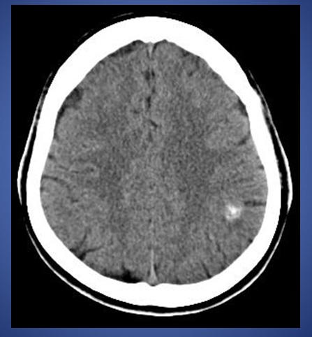
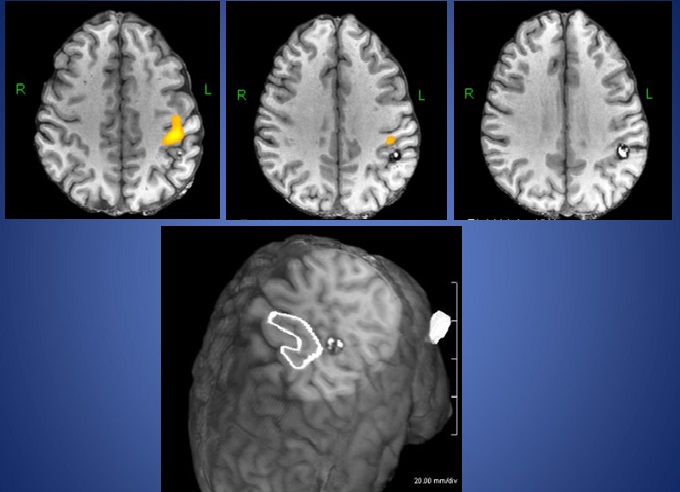
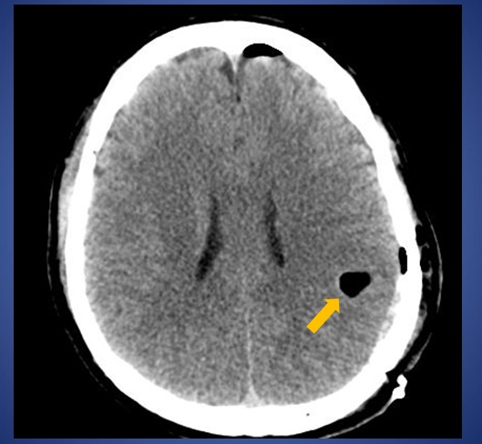
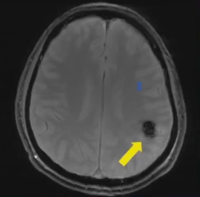
Cavernous Malformations. For help with symptoms, pain, risks, and treatment contact us or see Seattle Neurosciences’ website.

Dr. Newell was born in Boston, MA and attended Case Western Reserve University medical school. He then completed his residency in neurosurgery at the University of Washington, including one year in London at St. George’s medical school. Dr. Newell is the co-founder of the Swedish Neuroscience Institute and founder of the Seattle Neuroscience Institute.